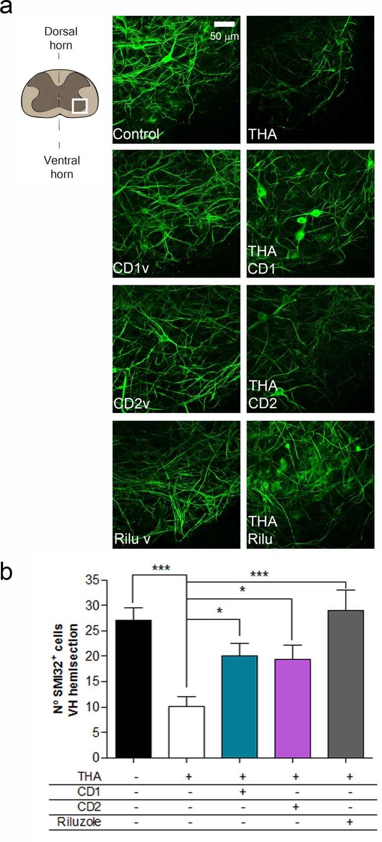
Fig 2. In silico design of polypharmacology for neuroprotection in an in vitro model of ALS.

a. Left, Schematic drawing indicating the site of analysis (white frame) of MN survival at the ventral horn of the spinal cord slice. Middle and right, Representative microphotographs of MNs in the ventral horn of the spinal cord slice detected by immunohistochemistry with the SMI-32 antibody at 4 weeks after THA treatment. Mid panels show control culture and with addition of vehicle (v) for each drug combination (CD1-CD2) or riluzole. Right panels show cultures subjected to excitotoxicity by THA alone or with co-treatment with CD1 and CD2 drug combinations or riluzole. Scale bar, 50 μm. b. Bar graph showing the number (mean±SEM, n = 5) of SMI-32 positive cells in the ventral horn of each spinal cord slice. (***p<0.001; *p<0.05 by Dunnett’s post-hoc test vs THA condition).
