
Fig 5. Lack of anti-inflammatory effect exerted by single drugs.
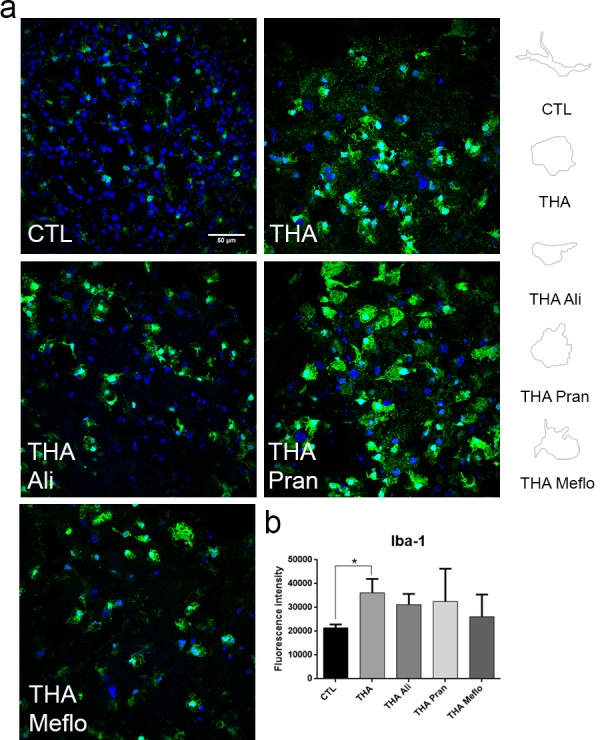
a. Representative microphotograhs of microglia and DAPI at 3 weeks after co-treatment with THA alone or plus single components of the neuroprotective combinations (CD1 and CD2): Aliretinoin (Ali), Mefloquine (Meflo) and Pranlukast (Pran) (n = 5). Right panels are profile drawings of microglia showing the ameboid-like or ramified shape acquired after each treatment. b. Bar graph showing the microglial reactivity of each experimental condition by measuring the immunofluorescence intensity of Iba-1 in the ventral horn of each spinal cord slice. (mean±SEM, n = 5) (*p<0.05; by Dunnett’s post-hoc test vs THA condition).