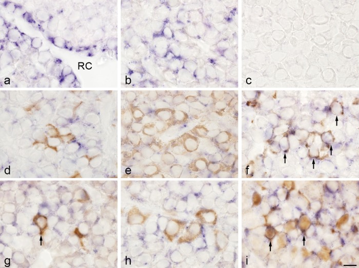
Fig. 3. .
Double-staining of TIMP2 mRNA detected by in situ hybridization and hormones and S100 protein detected by immunohistochemistry in rat anterior pituitary gland. a, b: In situ hybridization for TIMP2. TIMP2-expressing cells were observed in the marginal layer surrounding Rathke’s cleft (RC) (a) and in the anterior lobe (b). c: Negative control with sense probe. d–i: In situ hybridization of TIMP2 and immunohistochemistry of ACTH (d), GH (e), prolactin (f), TSHβ (g), LHβ (h), and S100 protein (folliculostellate cells; i). In situ hybridization with NBT/BCIP (blue) and immunostaining with 3,3'-diaminobenzidine (brown). TIMP2 mRNA was colocalized with the prolactin, TSHβ, and S100 protein immunoreactions (arrows). Bar=10 μm (a–i).

