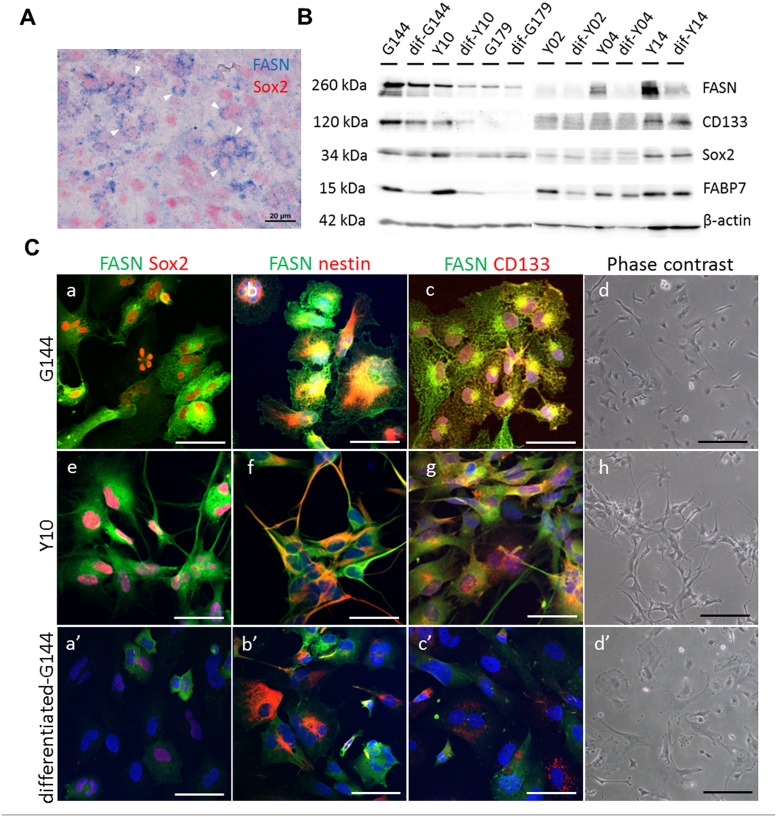
Fig 2. Expression of FASN in human glioblastoma cells and GSC lines.
(A) Human glioblastoma cells show the strong expression of FASN (blue) and the neural stem cell marker, Sox2 (red). Sox2 expression is confined to the nuclei, whereas FASN is expressed in the cytosol. The white arrows show FASN+Sox2+ cells. (B) Western blotting showing the expression of FASN, CD133, Sox2, and FABP7 in G144, Y10, G179, Y02, Y04 and Y14 GSC lines. Upon differentiation in the presence of FBS, FASN expression, similar to that of CD133, Sox2, and FABP7, was down-regulated. Expression of β-actin was used as an internal control. (C) Expression of FASN in GSC lines before and after differentiation. GSCs show strong expression of FASN and other neural stem cell markers, Sox2, nestin and CD133. Upon differentiation in the presence of FBS, FASN expression is down-regulated, similar to that of Sox2, nestin, and CD133. Immunofluorescence micrographs showing the co-expression of FASN with Sox2 (a, e, a’) and nestin (b, f, b’) in G144 and Y10 GSC lines. Phase contrast micrographs showing the morphology of GSC lines (G144 and Y10) in the presence of EGF and FGF (d, h) or after differentiation in the presence of FBS (d’). Bars in a-c, e-g, a’-c’ = 50 μm, Bars in d, h, d’ = 20 μm.

