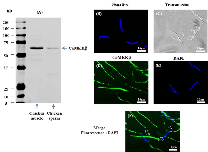
Fig 2. Presence and localization of CaMKKβ in chicken sperm.
Chicken sperm lysates (45μg of protein) were analyzed by western blotting using anti- CaMKKβ as primary antibody. Cell lysates from chicken muscle (40μg of protein) were used as positive control. A band of approximately 65kDa for CaMKKβ was detected (2A). Indirect immunofluorescence of chicken sperm was carried out with the same antibody. Negative control: primary antibody was not added (2B). White arrows and circles in transmission images indicate areas of the acrosome (arrow 1), the nuclei (arrow 2), the midpiece (circle), and the principal piece of the flagellum (arrow 3) in sperm (2C). Immunofluorescence staining of CaMKKβ (2D, green) was conducted; nuclei were stained with DAPI (2E, blue). Merged images of fluorescence with DAPI staining are shown in Fig 2F (white arrows indicate regions containing CaMKKβ immunoreactivity: arrow 1: acrosome; arrow 3: principle piece; circle: midpiece). Scale bar: 10μm.

