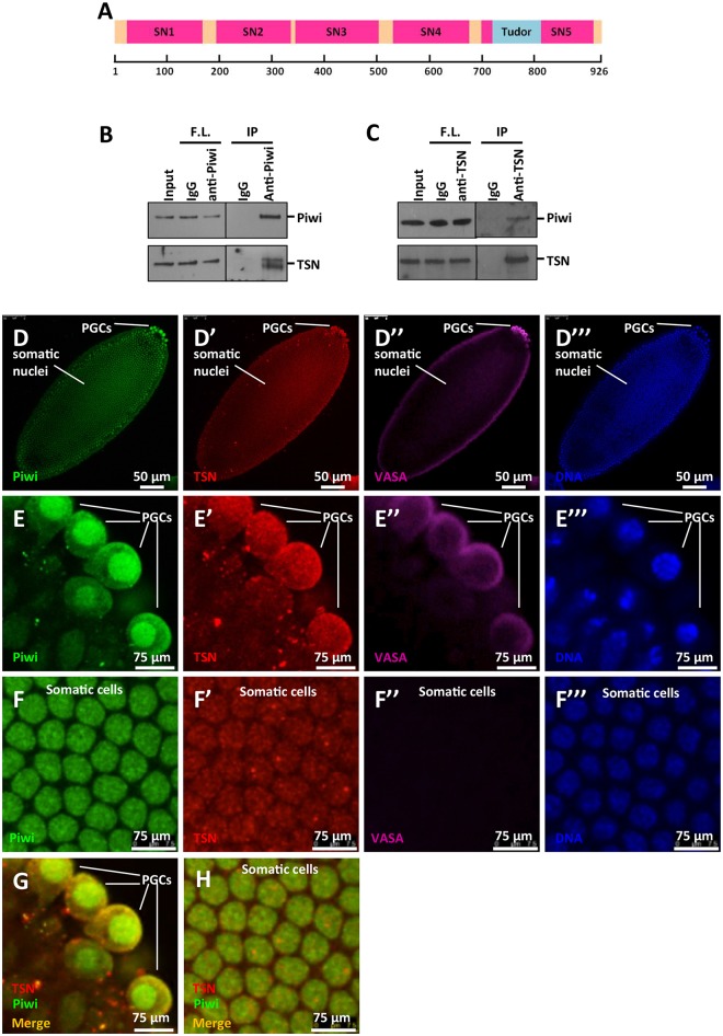
Fig 1. TSN is a novel Piwi-interacting protein.
(A) Schematic depiction of protein domain structures of TSN. TSN contains five staphylococcal nuclease-like domains (SN1-SN5) and a methyl lysine/arginine recognizing Tudor domain. (B) Co-immunoprecipitations of Piwi and TSN with anti-Piwi antibody. Input was the cytoplasmic fraction of the lysates from 0-12h WT embryos. 5% of input was used in the Western blot analysis. IgG was used as the control. F.L., flow-through; IP, immunoprecipitates. (C) Reciprocal co-immunoprecipitations of TSN and Piwi with mouse anti-TSN antibody. Input was the cytoplasmic fraction of lysates from 0–12 h WT embryos. 10% of input was used in the Western blot analysis. IgG was used as controls for the co-immunoprecipitations. F.L., flow-through; IP, immunoprecipitates. (D-F‴) Immunostaining of Piwi (green), TSN (using rabbit anti-TSN-N antibody, red), and the germ cell marker VASA (purple) in 0-1h WT embryos. DNA was labeled by DAPI (blue). (D-D‴) The whole embryo. PGCs, primordial germ cells. (E-E‴) Magnified images of E-E‴ showing the region of PGCs. Piwi and TSN were both highly enriched in the cytoplasmic region of PGCs. (F-F‴) Magnified images of D-D‴ showing the somatic cells. Piwi and TSN were both localized to the somatic nuclei. (G) Co-immunostaining of Piwi and TSN in PGCs, merged image of E and E’. (H) Co-immunostaining of Piwi and TSN in somatic cells, merged image of F and F’.

