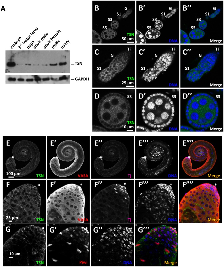
Fig 2. TSN is highly expressed in embryos and adult gonads.
(A) Expression pattern of TSN protein in different key developmental stages and tissues. Western blot analysis using mouse anti-TSN antibody revealed that TSN was highly expressed in the embryonic stage as well as adult ovaries and testes. (B-D”) Immunostaining of TSN (using mouse anti-TSN antibody, green) in adult WT ovaries. DNA was labeled by DAPI (blue). TSN was localized to the cytoplasm of both germline and somatic cells in adult ovaries. G, germarium; S1-S5, stage 1–5 egg chambers, respectively; TF, terminal filament. (C-C”) Magnified images of a germarium including a S1 egg chamber. (D-D”) Magnified images of B-B” showing the S3 egg chamber. (E-F””) Immunostaining TSN (using mouse anti-TSN antibody, green) in WT adult testes. Germ cells were labeled with anti-VASA antibody (red) and somatic cells were labeled with anti-Tj antibody (purple). DNA was labeled by DAPI (blue). TSN was localized to the cytoplasm of germline and somatic cells in adult testes. Magnified images of the apical region of the testis are shown in F-F””. (G-G‴) Immunostaining of TSN (green) and Piwi (red) in WT adult testes. DNA was labeled by DAPI (blue). Piwi was localized in the nuclei of hub cells, early germ cells and somatic cyst cells. Asterisk: the hub.

