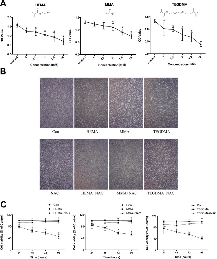
Fig 1. Dental monomers induced cytotoxic effects in hDPCs.
A. Cell viability of hDPCs after dental monomer treatment (HEMA: 1–2.5-5-7.5–10 mM; MMA; 1–2.5–5–7.5–10 mM; or TEGDMA: 1–2.5-5-7.5–10 mM) for 24 h, as analyzed by the CCK-8 assay. B. Morphological changes in hDPCs after treatment with dental monomers (1mM HEMA, 5mM MMA or 1mM TEGDMA) in the absence or presence of 10 mM NAC. C. Cell viability of hDPCs after exposure to dental monomers (1mM HEMA, 5mM MMA or 1mM TEGDMA) without or with 10 mM NAC. Data represent the mean ± SD of three independent experiments (n = 6). *P < 0.05 vs. control group by one-way ANOVA.

