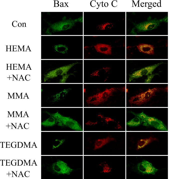
Fig 7. Double Immunofluorescence staining for Bax and Cyto C in hDPCs exposed to dental monomers in the absence or presence of NAC.
Green fluorescence represents Bax, whereas red green fluorescence represents Cyto C. Yellow color in the overlay of these two images indicates co-localization of Bax and Cyto C (presumably in mitochondria).

