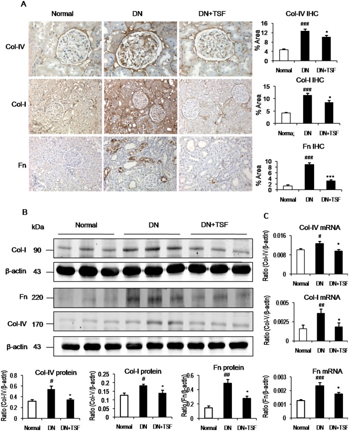
Fig 3. TSF inhibits renal fibrosis in DN rats.
(A) Immunohistochemistry (IHC) and quantitative analysis of collagen IV (Col-IV, original magnification, × 400), collagen I (Col-I, original magnification, × 200), and fibronectin (Fn, original magnification, × 200). (B) Western blot and quantitative analysis of collagen I, collagen IV, and fibronectin. (C) Real-time PCR of collagen I, collagen IV, and fibronectin. Results show that compared with the normal rats, renal fibrosisis markedly enhanced in DN rats. However, in TSF-treated DN rats renal fibrosis is inhibited as demonstrated by Western blot analysis, IHC at the protein level and real-time PCR at the mRNA level. Data are expressed as means ± SE for each group of 9 rats. *P<0.05, **P<0.01 TSF-treated group vs. DN group; #P< 0.05, ##P<0.01, ###P<0.001 DN group vs normal group.

