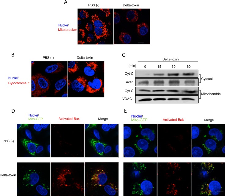
Fig 7. Delta-toxin caused mitochondrial dysfunction.
(A) A549 cells preincubated with MitoTracker red and Hoechst 33342 were treated with delta-toxin (50 ng/ml) for 30 min at 37°C. (B) A549 cells were treated with delta-toxin (50 ng/ml) for 30 min at 37°C. Cells were formaldehyde-fixed, permeabilized and stained using an anti-cytochrome c antibody and Hoechst 33342. (C) A549 cells were treated with delta-toxin (50 ng/ml) for the indicated time periods at 37°C. The mitochondria and the cytosol fractions were prepared as described in the Materials and Methods, and then were subjected to immunoblotting for the detection of cytochorome c. (D,E) A549 cells transfected with Mito-GFP were treated with delta-toxin (50 ng/ml) for 30 min at 37°C. The cells were formaldehyde-fixed, permeabilized and stained with an active-form-specific anti-Bax antibody (D) or an active-form-specific anti-Bak antibody (E). Nuclear DNA was stained with Hoechst 33342. Cells were examined using a confocal microscope. These results are representative of four experimental studies. Bar, 5 μm.

