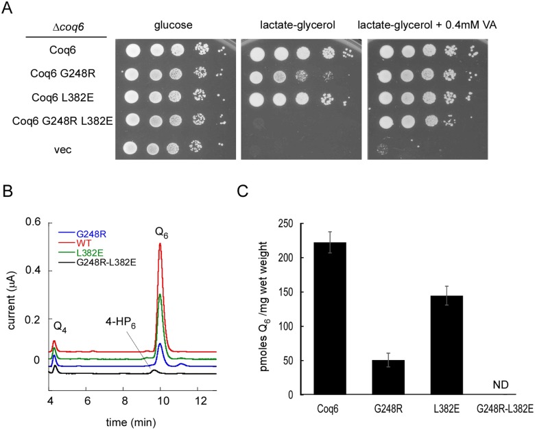
Fig 10.
A) 10 fold serial dilution of the Δcoq6 strain carrying an empty plasmid (vec) or plasmids coding for Coq6p, Coq6p-G248R, Coq6p-L382E and Coq6p-G248R-L382E. The plates contained YNB-pABA agar medium supplemented with the indicated carbon source and vanillic acid (VA) or not. The plates were imaged after incubation at 30°C for 2 days (glucose) or 6 days (lactate-glycerol). B) Representative electrochromatogram of lipid extracts from Δcoq6 cells expressing either Coq6p, Coq6p-G248R, Coq6p-L382E and Coq6p-G248R-L382E (1 mg of cells). The elution position of the Q4 standard, of 3-hexaprenyl-4-hydroxyphenol (4-HP6) and Q6 are indicated. C) Q6 amounts (in pmoles per mg of wet weight) in Δcoq6 cells expressing either Coq6p, Coq6p-G248R, Coq6p-L382E or Coq6p-G248R-L382E. Cells were grown in YNB–pABA 2% lactate-glycerol containing 10 μM 4HB. The results are the average of 3–4 independent experiments and error bars represent standard deviation. ND, not detected.

