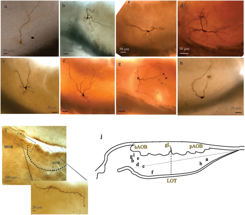
Figure 4.
Large principal cells of anterior and posterior halves of the accessory olfactory bulb (AOB). (A–I) Representative pictures of recorded AOB-large principal cells (LPCs). Note the variable morphology of this population of principal cells and their distinct degrees of glomerular innervation. Neuron in i is a “rhythmic” neuron, note its far reaching dendrite leaving the AOB. (J) Camera lucida drawing showing the approximate position of the neuron's somata.

