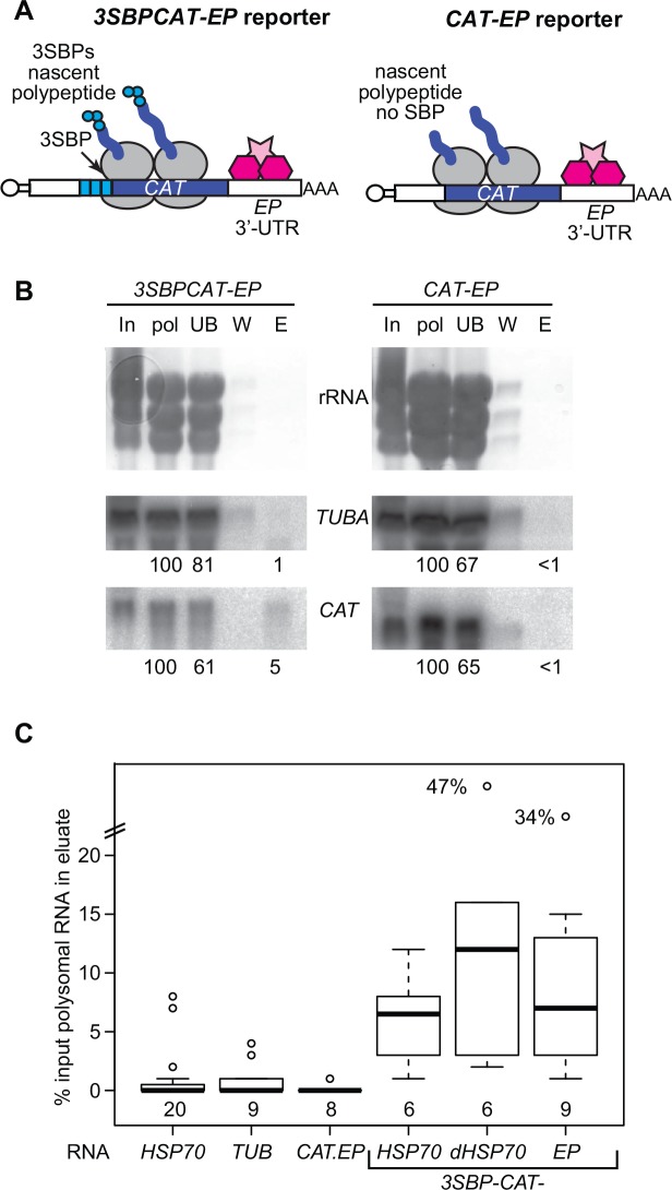
Fig 2. Purification of the 3SBP-CAT-SKL-EP mRNP.
(A) The mRNAs. Multi-tag reporter (upper left) composed of 3SBPs at the N-terminus of the ORF and the control reporter (upper right), without the 3SBPs. SBP: streptavidin binding peptide; UTR: untranslated region; CAT: chloramphenicol acetyltransferase; SL: spliced leader; RBP: RNA-binding protein. The black portion is the CAT gene. (B) Northern blot showing purification of the 3SBP-CAT-SKL-EP mRNA and failure to purify CAT-SKL-EP mRNA. TUBA: alpha tubulin. The numbers below the blots are the relative amounts of the mRNA measured, relative to the polysomal RNA input. These numbers are already correcting for loading. In: input cells (6x10⁷ cell-equivalents): pol: polysomes (6x10⁷ cell-equivalents); UB: unbound (6x10⁷ cell-equivalents); W: wash (6x10⁷ cell-equivalents); E: eluate (8x10⁷ cell-equivalents). Each probe detects a single band. (C) Box plot for all purifications similar to those in this Figure and Fig 3. The centre line is the median, the boxes extend over the 25th to 75th percentiles, and the whiskers show the 95% confidence limits. The number of independent experiments for each construct is shown beneath the boxes.

