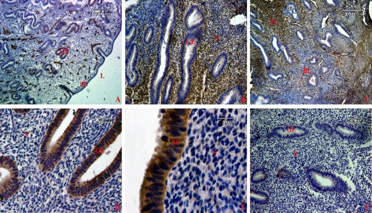
Fig. 2.
Localization of stem cell markers in human endometrial tissue samples. The brown color shows the expression of each marker by immunohistochemistry using diaminobenzidine as the substrate. A group of CD146 (A), CD90- (B) and CD105- (C) positive cells were localized in both basalis and functionalis stroma. The expression of CD9 (D) and EpCAM (E) were detected in epithelial cells. (F) Sections of endometrium were stained with secondary antibody- (without primary antibody) serving as a negative control in our study. S, stroma; SE, surface epithelium; GE, glandular epithelium; BL, basalis layer; FL, functionalis layer; L, lumen

