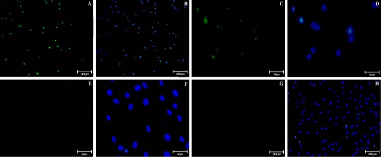
Fig. 6.
Immunostaining of pluripotency markers in cultured endometrial cells at fourth passage. Positive staining for Oct4 (A and B) and Nanog (C and D) markers were detected in endometrial cells. No staining were seen for Sox2 (E and F) and Klf4 (G and H) markers. The second and fourth column of the fluorescent photomicrographs represents the nuclei (blue colors: 6-diamidino-2-phenylindole) images from the same field of the immunofluorescence images

