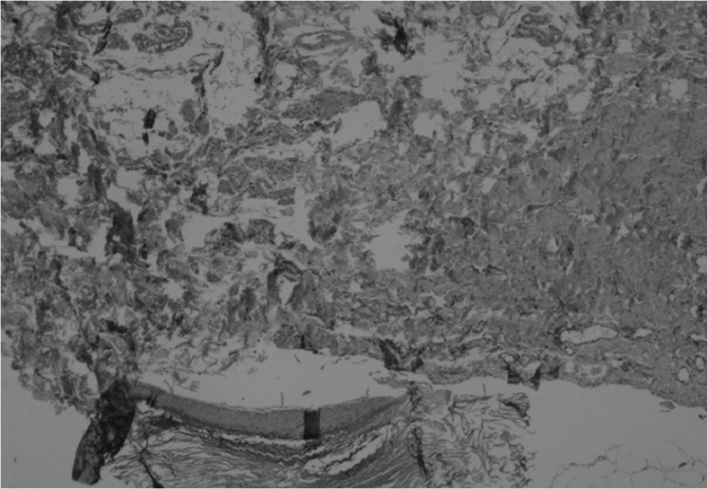
Figure 1.

Pathological analysis of an EIC of the breast as presented in a 70-year-old patient. Breast lobules may be observed in the upper section of the image, and the EIC may be observed in the lower section. Stain, hematoxylin and eosin; magnification, ×100; EIC, epidermal inclusion cyst.
