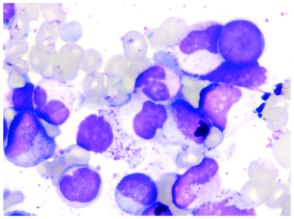
Figure 1.
Case one: Morphological examination of the bone marrow showing atypical chronic myeloid leukemia. Readily identifiable marrow blasts, a relatively high proportion of neutral promyelocytes displaying a pathological phenotype, and reduced cytoplasmic granules in the granulocytes are observed. Wright's stain; magnification, ×100.

