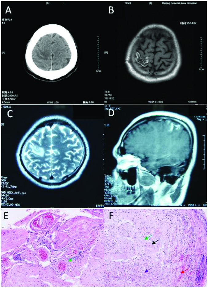
Figure 3.
Neuroimaging and pathological studies for patient 3 show evidence of isolated cortical vein thrombosis. (A) Brain computed tomography scans show hypodense lesion with some hyperdense within the right parietal lobe. (B) Brain axial magnetic resonance imaging (MRI) shows long signal with short signal in the right parietal lobe in the T1-weighted image. (C) Brain axial MRI shows long signal in the right parietal lobe in the T2-weighted image. (D) Partial focus was enhanced in the contrast-enhanced MRI. (E and F) Histopathologic analysis was sporadic hemorrhage (red arrow), tissue structure disappearance, astrocytic reaction and microglia (purple arrow), proliferation of small vessels (black arrow) and perivascular cuffing (green arrow). Amplification of G was 400 times and H was 200 times.

