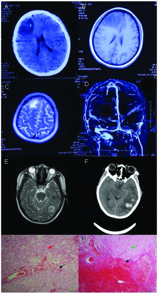
Figure 4.
Neuroimaging and pathological studies for patient 4 show evidence of isolated cortical vein thrombosis. (A) Brain computed tomography (CT) scan shows hypodense lesion with hyperdensity within the right frontal lobe. (B) Brain magnetic resonance imaging (MRI) shows long signal with short T1 signal in the right frontal lobe. (C) Brain MRI shows long T2-weighted image in the right frontal lobe. (D) In the brain MRV, the major venous sinuses were patent and did not show any abnormal signals. (E) Brain MRI shows long T2-weighted image in the left temporal lobe. (F) Brain CT scan shows hypodense lesion with hyperdensity within the left temporal lobe. (G and H) Histopathologic analysis revealed focal necrosis, and hemorrhage with lots of gitter cells (red arrow). Histopathologic analysis revealed astrocytic reaction, degeneration of neurons and endothelial proliferation in small vessels (green arrow). Histopathologic analysis revealed focal vascular mass with vasodilatation and congestion (black arrow). Amplification of G and H was 200 times.

