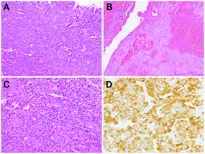
Figure 2.
Histopathological features of the epidural lesion. (A) The lesion was a highly cellular lesion composed of epithelial cells (H&E stain; original magnification, ×100). (B) The lesion was accompanied by organizing blood clots (H&E stain; original magnification, ×100). (C) The tumor cells exhibited abundant eosinophilic cytoplasm and a trabecular growth pattern consistent with metastatic hepatocellular carcinoma (H&E stain; original magnification, ×200). (D) The tumor cells were markedly positive for Hep Par 1 expression (immunohistochemistry; original magnification, ×400). H&E, hematoxylin and eosin.

