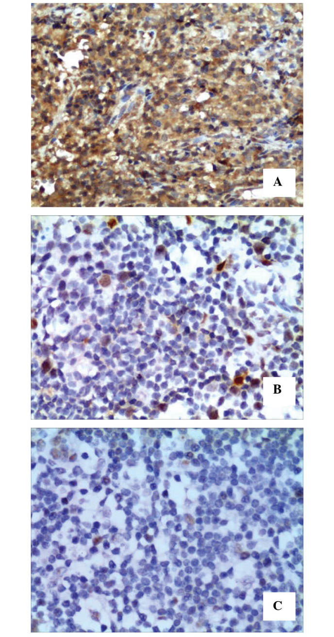
Figure 2.

Immunohistochemical staining of TIAM1 in clinical FFPE tissues (magnification, ×200). The rate of positive cells and the intensity of the staining for TIAM1 protein was higher in (A) extranodal natural killer/T-cell lymphoma, nasal type FFPE tissues, compared with (B) reactive lymph node hyperplasia and (C) normal lymph node tissues. TIAM1, T-lymphoma invasion and metastasis inducing factor 1; FFPE, formalin-fixed paraffin-embedded.
