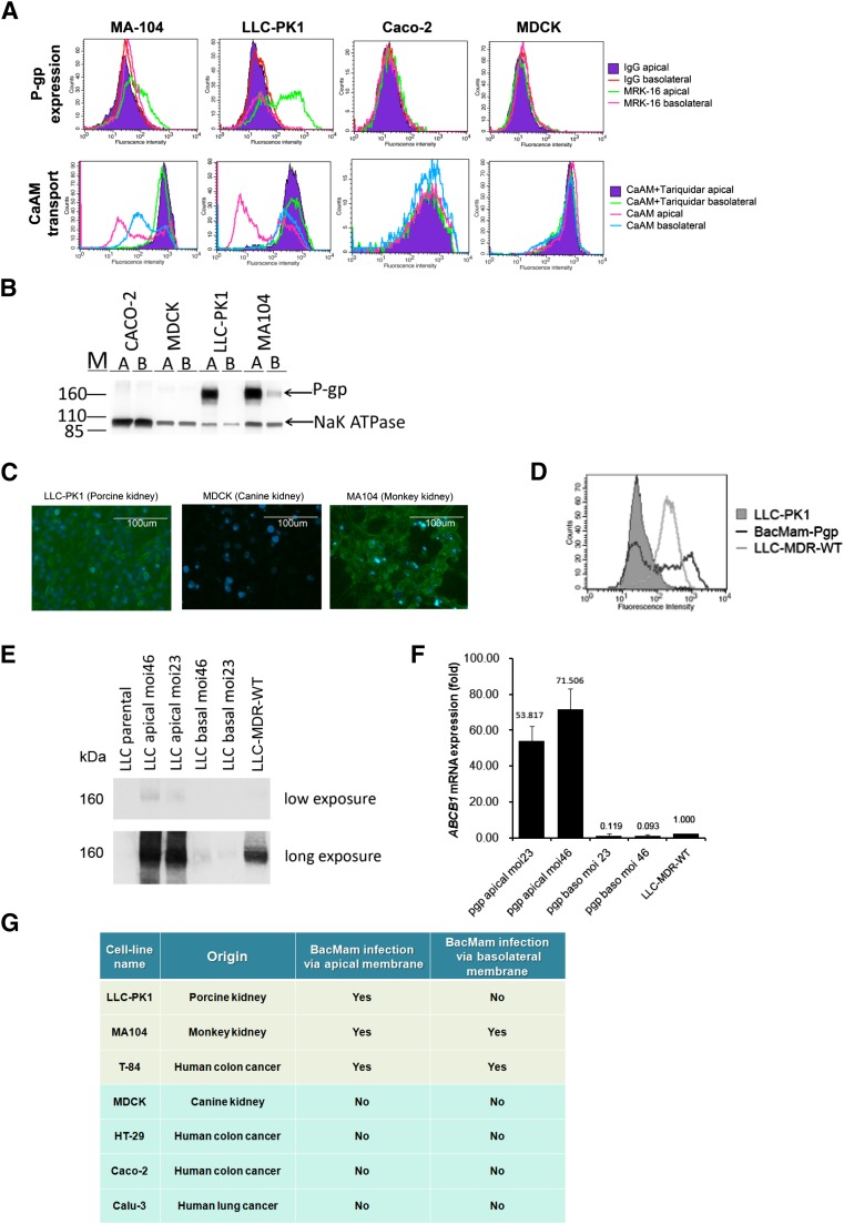
Fig. 1.
LLC-PK1 and MA104 monolayers express human P-gp upon transduction with BacMam baculovirus. (A) Flow cytometry histograms showing recombinant P-gp expression and ability to transport calcein-AM (CaAM) in the presence and absence of TQR (1 μM) in MA104, LLC-PK1, Caco-2, and MDCK cell lines. (B) Western blot analysis showing recombinant P-gp expression in Caco-2, MDCK, LLC-PK1, and MA104 cells. BacMam-Pgp virus was transduced to the polarized cells via the apical membrane [A] and via the basolateral membranes [B]. Na+/K+- ATPase was used as a loading control. (C) Microscopic images showing recombinant GFP expression in LLC-PK1, MDCK, and MA104 cell monolayers. Nuclear staining by DAPI (blue) in each cell line is also shown. (D) Flow cytometry histograms showing P-gp expression levels in BacMam-Pgp–transduced LLC-PK1 cells (black line) and LLC-MDR-WT cells (gray line). LLC-PK1 cells were used as a control (gray filled histogram). (E) Expression of recombinant P-gp by BacMam is influenced by BacMam virus to cell ratio. LLC-PK1 cells were transduced with BacMam virus on the apical and basolateral membranes with MOI 23 and MOI 46. Western blot analysis showing the level of P-gp expression. Short and long exposures are shown. LLC-PK1 cells and LLC-MDR-WT cells were used as negative and positive controls, respectively. (F) Exogenous ABCB1 mRNA expression is influenced by BacMam virus concentration. Bar graph showing the expression of recombinant human P-gp in the BacMam transduced cells relative to the LLC-MDR-WT cells. Glyceraldehyde-3-phosphate dehydrogenase was used as an endogenous gene control. (G) Table summarizing cell monolayers susceptible to transduction with BacMam baculovirus.

