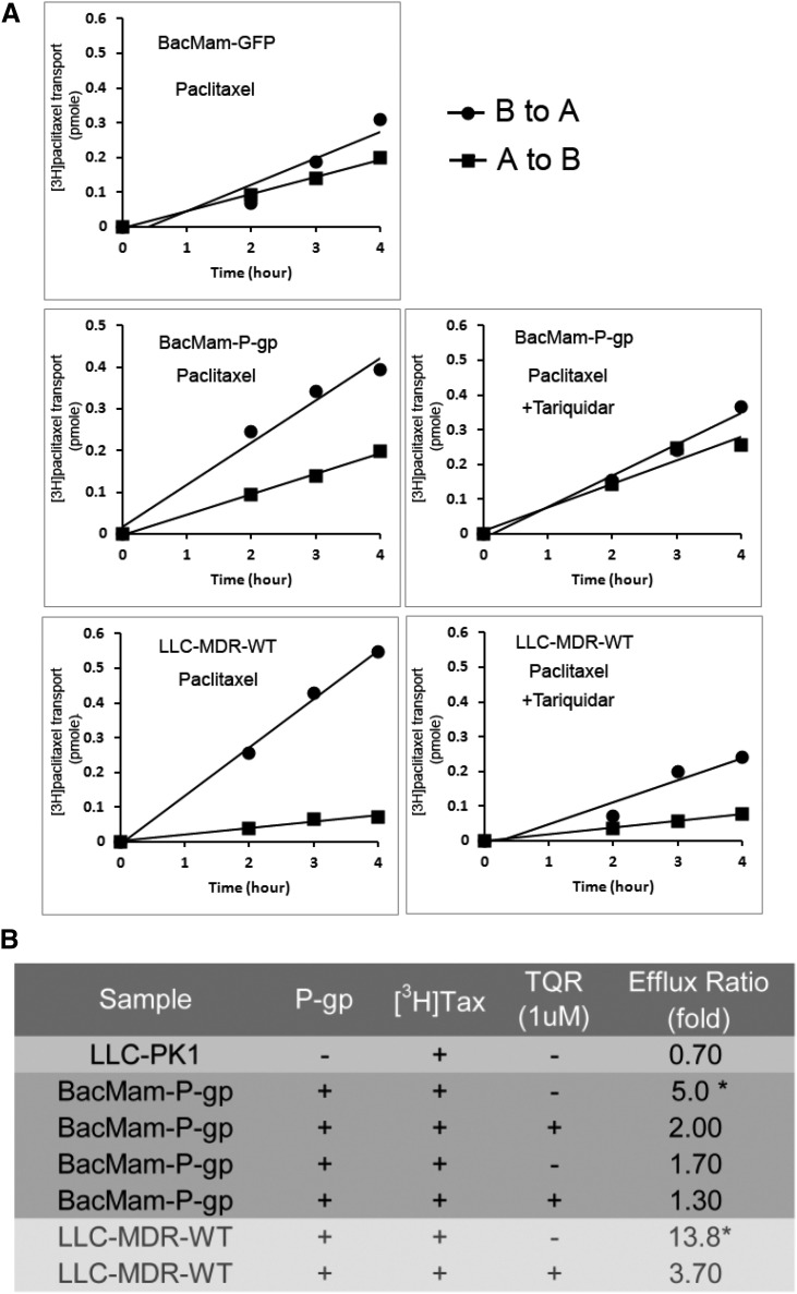
Fig. 3.
Transepithelial [3H]-paclitaxel transport across LLC-MDR-WT and BacMam-P-gp– or BacMam-GFP–infected LLC-PK1 cells. (A) Plots showing time-dependent increase of [3H]-paclitaxel on the receiver side of the transwells with cells transduced by BacMam-GFP or BacMam-P-gp. LLC-MDR-WT cells were used to compare [3H]-paclitaxel vectorial transport. To inhibit P-gp efflux activity, 1 μM TQR was added 10 minutes before adding [3H]-paclitaxel. Each point is the mean ± S.D. (n = 3). (B) Table summarizes the fold of [3H]-paclitaxel transport across LLC-PK1 cells, LLC-PK1 cells transduced with BacMam-P-gp and LLC-MDR-WT cells. To inhibit P-gp function, 1 μM TQR was added. Transport of [3H]-paclitaxel was calculated as efflux ratio in terms of fold of drug transport (B→A/A→B) in a period of 4 hours. *P < 0.5.

