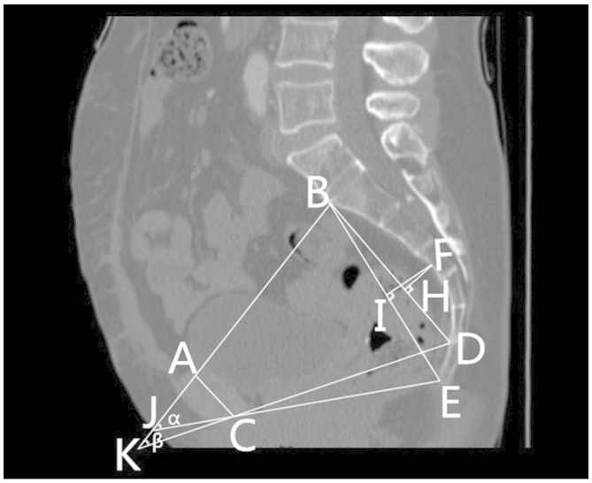
Figure 2.
Mid-sagittal view of the pelvis in a female patient, indicating the (FI) depth of the sacrococcygeal curvature, (FH) the depth of the sacral curvature and the (α) sacrococcygeal-pubic and (β) sacropubic pelvic angles. (A) The superior median aspect of the pubic symphysis. (B) The anterior median aspect of the sacral promontory. (C) The inferior median aspect of the pubic symphysis. (D) The anterior median aspect of the sacrococcygeal junction. (E) The inferior median aspect of the tip of the coccyx. (F) The deepest portion of the sacral hollow or sacrococcygeal hollow. (H) A point of the perpendicular line from the deepest portion of the sacral hollow to the sacral distance line. (I) A point of the perpendicular line from the deepest portion of the sacrococcygeal hollow to the sacrococcygeal distance. (J) The point between an extension of the line forming the anteroposterior diameter of the pelvic inlet and that of the anteroposterior diameter of the pelvic outlet. (K) The point between an extension of the line forming the anteroposterior diameter of the pelvic inlet and that of the anteroposterior diameter of the mid-pelvis.

