Fig. 2.
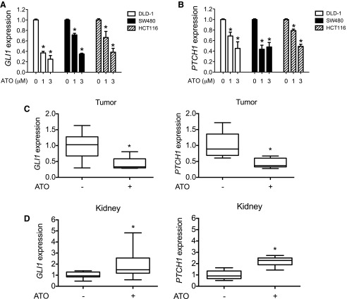
ATO modulates GLI signaling in a CRC xenograft mouse model. (A and B) CRC cells (DLD-1, SW480, and HCT116) were treated with the indicated concentrations of ATO for 48 hours, and GLI1 (A) or PTCH1 (B) expression relative to that of GAPDH was determined by qRT-PCR. (C and D) HCT116 cells (2 × 10⁶) were subcutaneously implanted into nude mice. When the tumor size reached approximately 200 mm3, 10 mg/kg ATO or vehicle control was injected for 2 consecutive days. Tumor (C) and kidney samples (D) were then harvested for RNA extraction and the expression of GLI1 and PTCH1 relative to that of GAPDH was determined by qRT-PCR. Error bars represent the S.D. (n = 10). *P < 0.05 calculated using a Wilcoxon signed-rank test. Normalized data are shown here because of the inherent difficulty in comparing mouse expression levels with human expression levels. GAPDH, glyceraldehyde-3-phosphate dehydrogenase.

