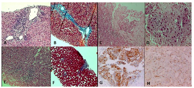
Fig. 1. - Histological images. A - Pretreatment liver biopsy: interface activity (hematoxylin-eosin, 100x). B - Pretreatment liver biopsy: fibrous septa (Masson's trichrome, 100x). C - Liver section from the tumor: moderately differentiated HCC (hematoxylin-eosin, 100 x). D - Liver section from the tumor: moderately differentiated HCC in detail (hematoxylin-eosin, 400x). E - Post treatment liver biopsy: non-tumoral tissue with mild activity (hematoxylin-eosin, 40 x). F - Post treatment liver biopsy: portal fibrosis (Masson's trichrome, 40x). G - HCC, immunohistochemistry: hepatocyte-specific antigen (400 x). H - HCC, immunohistochemistry: CD 10, canalicular staining (400 x).

