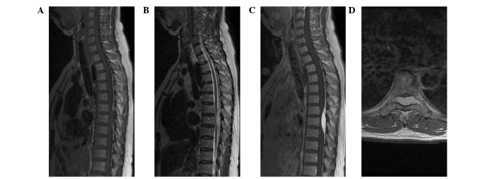
Figure 1.
Magnetic resonance imaging reveals a well-defined epidural tumor at the T6-T8 level. The tumor was isointense on T1-weighted imaging (WI) (A) and hyperintense on T2-WI (B). Marked homogeneous enhancement and the ‘dural tail sign’ were observed on contrast-enhanced T1-WI (C). Axial contrast-enhanced T1-WI revealed that the tumor was located dorsal-laterally, with severe cord compression and extension through the intervertebral foramen (D).

