
Figure 1.
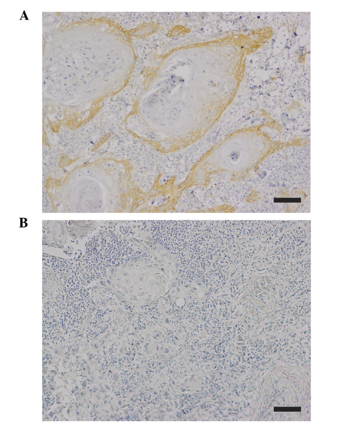
Immunohistochemical staining of EGFR in human OSCC tissues. (A) Positive immunoexpression of EGFR in OSCC. (B) Specificity of the staining was confirmed by using non-immune antibody instead of primary antibody as a negative control. Scale bar, 100 µm. EGFR, epidermal growth factor receptor; OSCC, oral squamous cell carcinoma.