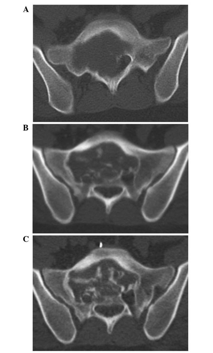
Figure 2.

CT imaging of the sacrum. (A) Sacral giant-cell tumor exhibiting an osteolytic lesion. (B) After 4 months of denosumab treatment, CT imaging of the sacrum revealed osteogenesis. (C) After 8 months of denosumab treatment, CT imaging revealed progression of osteogenesis, and preoperative vascular embolization was performed. CT, computed tomography.
