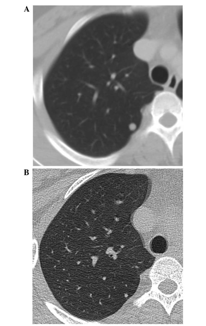
Figure 3.

(A) Enhanced chest CT imaging revealed a right pulmonary nodule measuring 6 mm in diameter, which was confirmed to be a metastatic lesion. (B) After 10 months of denosumab treatment, CT imaging revealed that the pulmonary nodule had reduced in size. CT, computed tomography.
