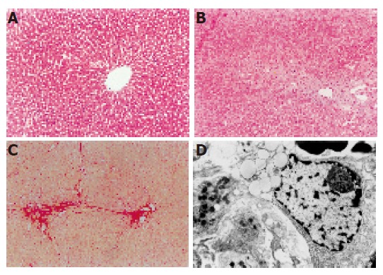
Figure 2.

Changes after DMN treatment. A: Livers in the control group showed normal lobular architecture with central veins; B: after 7 d of DMN treatment, extensive necrosis was observed in portal areas; C: after 14 d of DMN treatment, thin fibrotic septa were formed joining central areas; D: after 21 or 28 d of DMN treatment, thick intralobular septa were evident.
