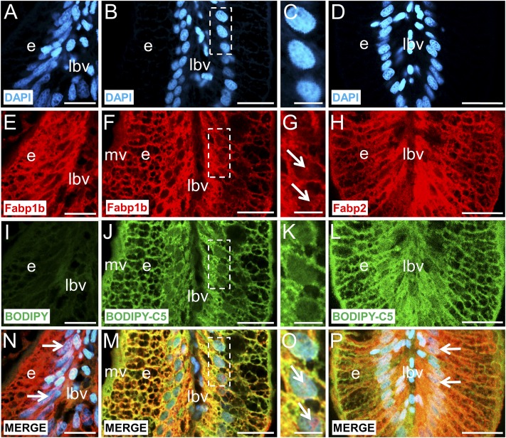
Fig. 9.
The intestinal absorption of dietary BODIPY-FLC5 and simultaneous localization of Fabp1b and Fabp2 in zebrafish intestinal villi. Histological sections were counterstained with DAPI to label the nuclei blue (A–D). The sections were stained with monospecific polyclonal antibodies developed against recombinant zebrafish Fabp1b.1 (E–G) or Fabp2 (H) proteins with Alexa 594-conjugated secondary antibody (red). Adult animals were previously fed with BODIPY493/503 mixed with CEY powder and used as a negative control (see Materials and Methods for details) (I) or BODIPY-FLC5 mixed with CEY powder (J–L), and fluorescence was detected on histological sections in green fluorescent protein channel (green). (N–P) Merging between nuclei labeling, immunofluorescence signal of Fabps, and BODIPY493/503 or BODIPY-FLC5 signals are presented for each corresponding column of the figure. The apex of the villi is positioned at the bottom of each image. C, G, K, and O are enlargements of the indicated area on B, F, J, and M, respectively. No BODIPY-FLC5 signal was detected inside the nuclei and the intranuclear overlap between Fabp and DAPI labels is shown as pink spots (white arrows in N, O, P). e, enterocyte; lbv, lymph and blood vessels; mv, microvilli. Scale bar represents 20 µm, except 5 µm in C, G, K, O.

