
Figure 3.
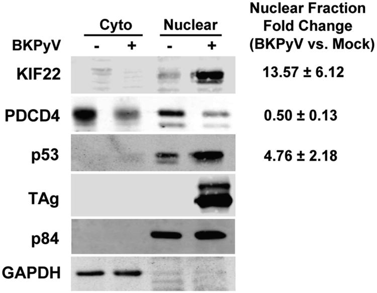
Western blotting validation of SILAC results. RPTEs were infected with BKPyV at an MOI of 0.5 IU/cell. At 3 dpi the nuclear and cytoplasmic fractions of the RPTEs were isolated and subjected to Western blotting analysis. Three protein targets were chosen from significantly up-regulated (p53 and KIF22) or down-regulated (PDCD4) proteins found in both SILAC experiments. p84 and GAPDH serve as fractionation markers for the nuclear and cytoplasmic fractions, respectively. Representative blots and quantitation using the Odyssey system (mean ± standard deviation) from three independent experiments were shown.