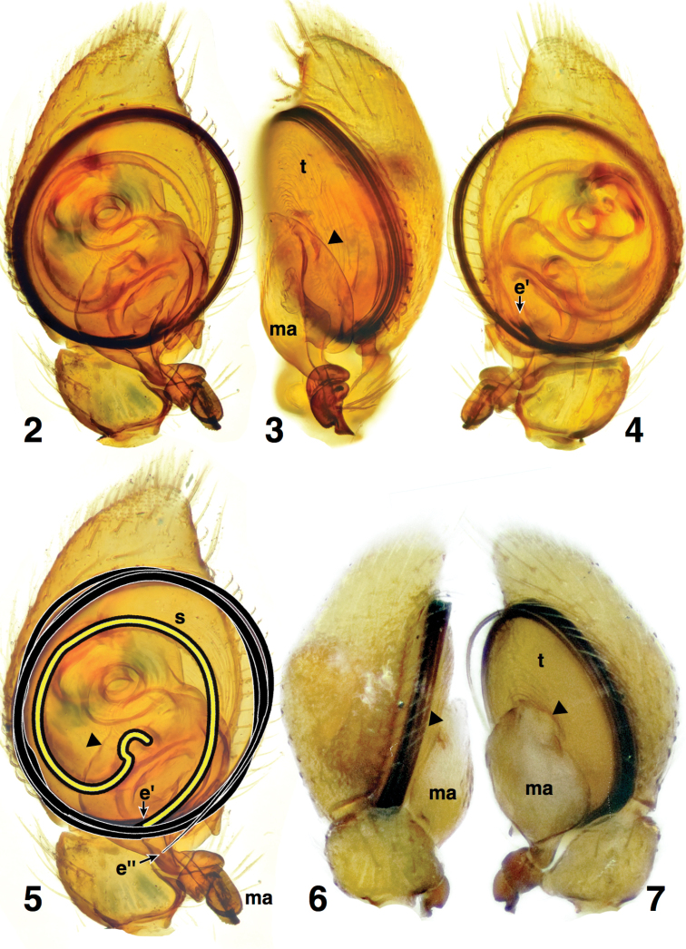
Figures 2–7.
Male palp of Depreissia myrmex 2 Cleared palp, ventral view. 3 Retrolateral view 4 Dorsal view 5 Ventral view, highlighting the path of spermaphore (yellow) and embolus (black) 6 Left palp, prolateral view 7 Right palp, prolateral view. Abbreviations: e’ = origin of embolus; e’’ = end of embolus; ma = median apophysis; s = spermophore; t = tegulum. Triangle marks base of median apophysis.

