
Figure 6.
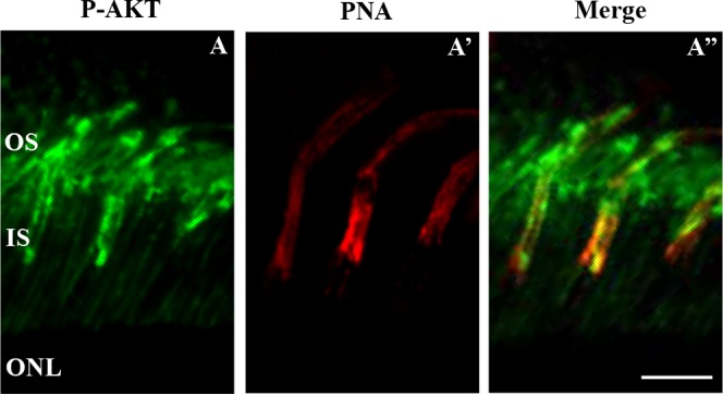
P-AKT is present in rods and cones. Fluorescence immunohistochemistry of C3H/f+/+ mice retinal sections at 3 months. Images were obtained at the level of the photoreceptor OS at ZT22. P-AKT (green) is seen in one specific structure (A). Cones are stained with PNA (red) while rods are unstained and visible as dark profiles (A'). In the merge, P-AKT is present in rod and cones OS (A''). Scale bar: 10 μm.