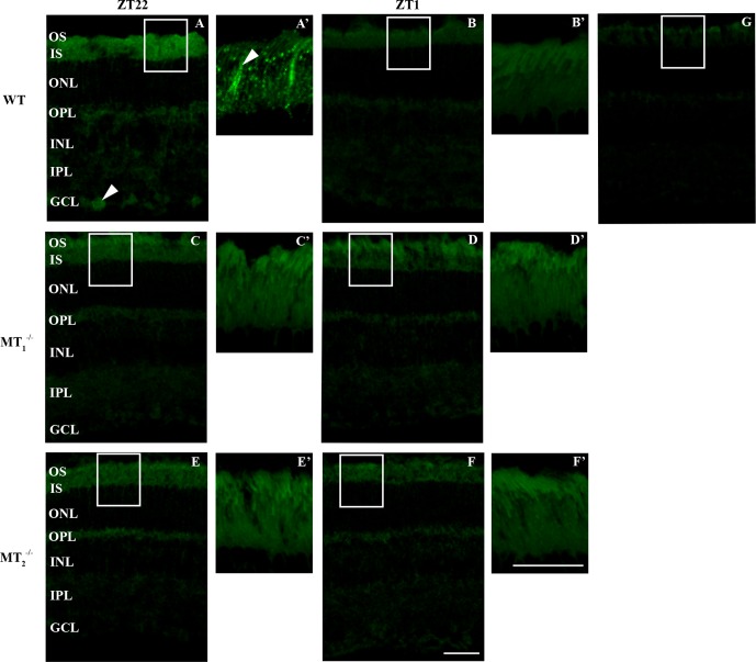
Figure 7.
P-FOXO1 is localized in photoreceptors and the GCL. P-FOXO1 localization by fluorescence immunohistochemistry performed on retinal sections of 3 months old C3H/f+/+, C3H/f+/+MT1−/−, and C3H/f+/+MT2−/− mice at ZT22 and ZT1. At ZT22, P-FOXO1 is localized in OS, IS and GCL (arrowhead) of C3H/f+/+ (A). Enlargement of boxed area shows the staining mainly in one specific structure in IS and OS (arrowhead, [A']). At ZT1, P-FOXO1 is not detected in the retina of C3H/f+/+ (B). Enlargement of the boxed area confirms no staining detected in OS and IS of C3H/f+/+ (B'). P-FOXO1 staining is very weak in OS, IS, and GCL of C3H/f+/+MT1−/− at ZT22 (C) and ZT1 (D) as in OS, IS, and GCL of C3H/f+/+MT2−/− mice at ZT22 (E) and ZT1 (F). Enlargement of boxed area confirms a very low level of P-FOXO1 in OS and IS of C3H/f+/+ MT1−/− and C3H/f+/+ MT2−/− mice (C'–F'). Control without primary antibody (G). Scale bars: 50 μm (A–F), 20 μm (A'–F').

