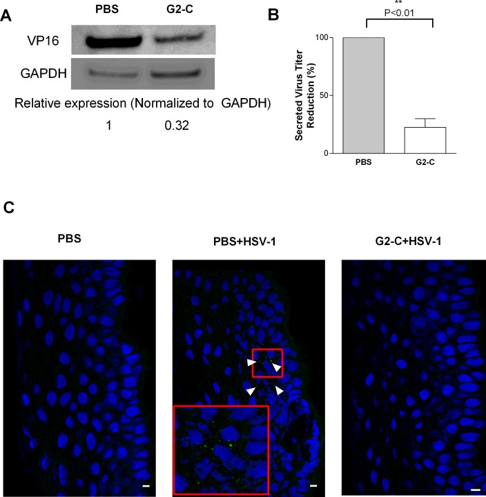
Figure 5.
Peptide G2-C released from the contact lens suppresses HSV-1 infection ex vivo. (A) Immunoblots representing the levels of the viral tegument protein VP16 from human cornea cadavers that were pretreated with the indicated treatments. Below: Band intensities were quantified using image analysis software (GE Healthcare) and were normalized to GAPDH. (B) The supernatant from the cultured human corneas were serially diluted and titered on Vero cells. Plaque numbers are normalized to PBS. Results are mean ± SEM of three independent experiments. Asterisks denote significant difference: **P < 0.01. (C) Representative sections from pig corneas that received the indicated treatments. The arrow indicates the presence of virus (green) on the epithelial cells (blue). Scale bars: 10 μm.

