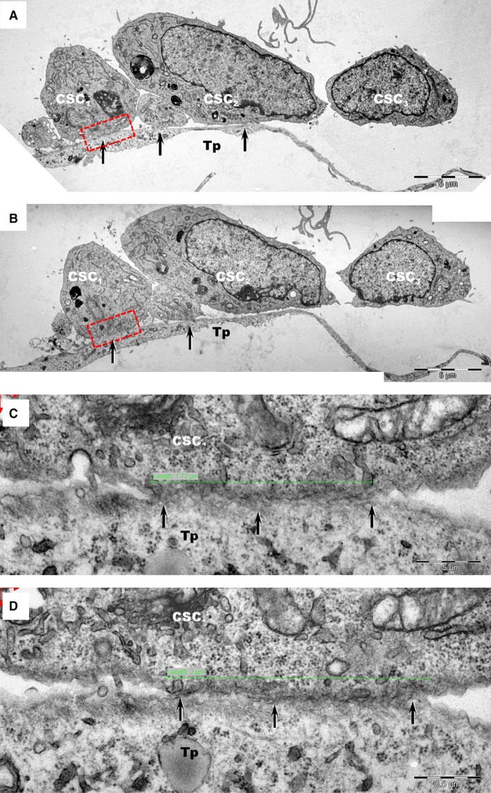
Figure 3.

Transmission electron microscopy images of TC–CSC culture after 24 hrs. (A, B) Serial sections show close contacts (black arrows) between cardiac stem cells (CSC 1‐CSC 3) and telopodes (Tp) of telocytes. (C, D) Higher magnification of rectangular marked areas in images A and B highlight the interface between cardiac stem cell CSC 1 and a telopode (Tp). An oblique sectioned stromal synapse (arrows) is visible between Tp and CSC 1. The length of the stromal synapse is about 2 μm (green line). Various types of vesicles may be seen at the interface between cells.
