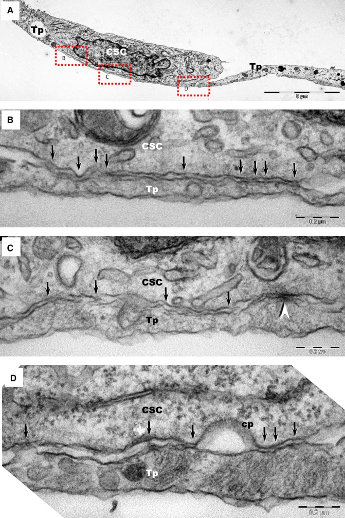
Figure 4.

(A) Transmission electron microscopy images of TC–CSC culture after 48 hrs shows a telopode (Tp) in close contact with a cardiac stem cell (CSC). (B, C, D) Marked areas from image A are shown at higher magnification in the corresponding panels. A planar contact (stromal synapse) between TC and CSC can be seen associated with a number of electron‐dense structures (arrows). A puncta adherentia junction (arrowhead) is visible between TC and CSC in image C. Cp – coated pit.
