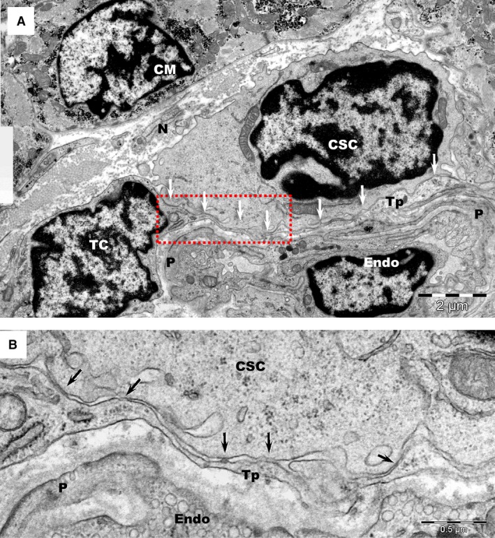
Figure 6.

(A) Transmission electron microscopy of human atrial tissue represents a glimpse into the complex environment of the cardiac stem cell niche, which comprises: telocytes (TC), cardiac stem cells (CSC), capillaries (Endo: endothelial cell; P: pericytes) and nerve endings (N). CM – adult cardiomyocyte. Arrows indicate the close contacts between a telopode (Tp) and a CSC. (B) Higher magnification of the rectangular marked area in image A.
