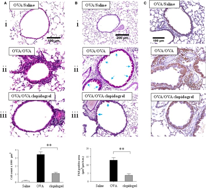
Figure 3.

The effect of clopidogrel treatment in lung tissue. (A) Lung tissue histology with haematoxylin and eosin and (B) periodic acid‐Schiff (PAS) staining. Quantitative analysis of inflammatory and PAS + cells in lung tissue was performed as described in the Materials and Methods section. (C) Immunohistochemistry of ECP was performed with an anti‐mouse ECP antibody. (i) Mice sensitized with OVA and challenged with saline (OVA/Saline vehicle); (ii) mice sensitized and challenged with OVA (OVA/OVA vehicle); (iii) OVA challenged mice treated with clopidogrel (OVA/OVA clopidogrel treatment). **P < 0.01 versus the OVA/OVA group.
