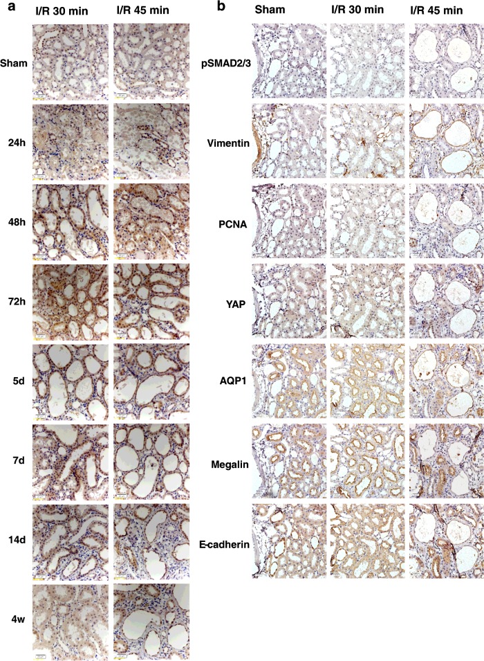
Figure 3. Immunohistochemical staining of YAP and immunostaining of serial sections of YAP co-expressed with differentiation markers.
(a) Immunostaining of YAP (×400 magnification) over time after I/R in 30 and 45 min groups. Scale bar, 20 μm. (b) Immunostaining of serial sections of YAP co-expressed with differentiation markers (×400 magnification) over time after I/R in 30 and 45 min groups. Scale bar, 20 μm.

