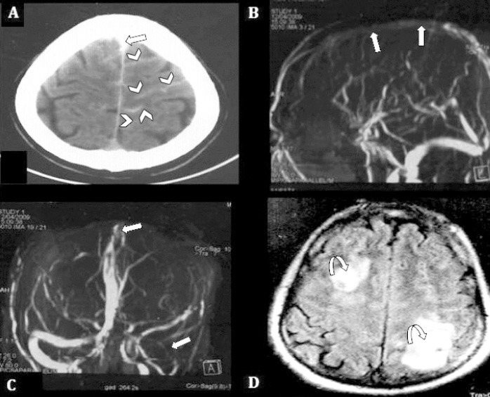
Figure 1.

Plain CT brain showing: A) SAH in paramedian sulci bilaterally over the left fronto-parietal region (cSAH), (arrow heads), and thrombosis of SSS (straight arrow). B) MRV 3D MIP image, anterior projection, demonstrate non-visualization of superior portion of SSS and draining veins. C) transverse and sigmoid sinus on the left side (straight arrows). D) FLAIR image, shows venous infarcts in frontal and parietal regions on both sides, (curved arrows). SAH - spontaneous subarachnoid hemorrhage, cSAH - convexity SAH, SSS - superior sagittal sinus, MRV - magnetic resonance venography, MIP - maximum intensity projection
