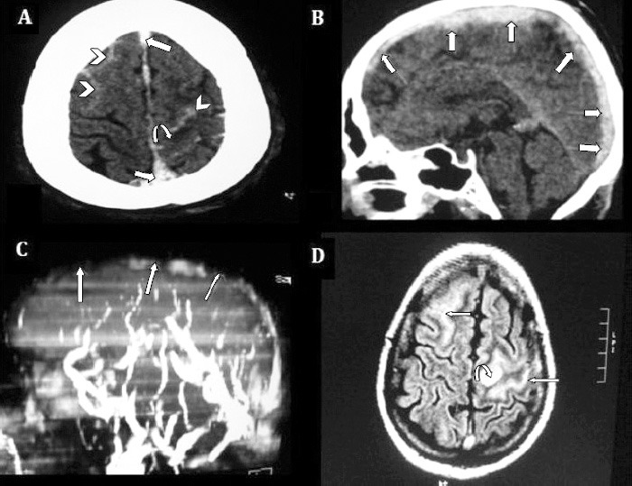
Figure 2.

Non-contrast enhanced axial CT brain showing: A) bilateral SAH (arrow heads), predominantly over left fronto-parietal sulci (cSAH) surrounded by hypodense venous infarct (curved arrow). B) Thrombosis of SSS also visualized in sagittal and axial cuts, (straight arrows). C) MRV 3D MIP image, left lateral projection, showed SSS thrombosis (straight arrows), with non visualization of its tributaries. D) FLAIR axial images demonstrate hyperacute SAH as hyperintense curvilinear signals in bilateral frontal lobes, (straight arrows), and hyperintense signals in frontal cortex consistent with venous infarct (curved arrow). SAH - spontaneous subarachnoid hemorrhage, cSAH - convexity SAH, SSS - superior sagittal sinus, MRV - magnetic resonance venography, MIP - maximum intensity projection
