Fig. 1.

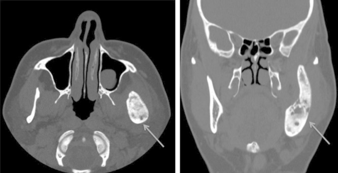
CT images. A and B show axial and coronal images, respectively, illustrating osteitic changes and thickening of the left hemimandible (arrows). Incidental note is made of a mucous retention cyst in the left maxillary sinus.

CT images. A and B show axial and coronal images, respectively, illustrating osteitic changes and thickening of the left hemimandible (arrows). Incidental note is made of a mucous retention cyst in the left maxillary sinus.