
Fig 1. Intranasal α-GalCer administration activates lung iNKT cells to secrete cytokines.
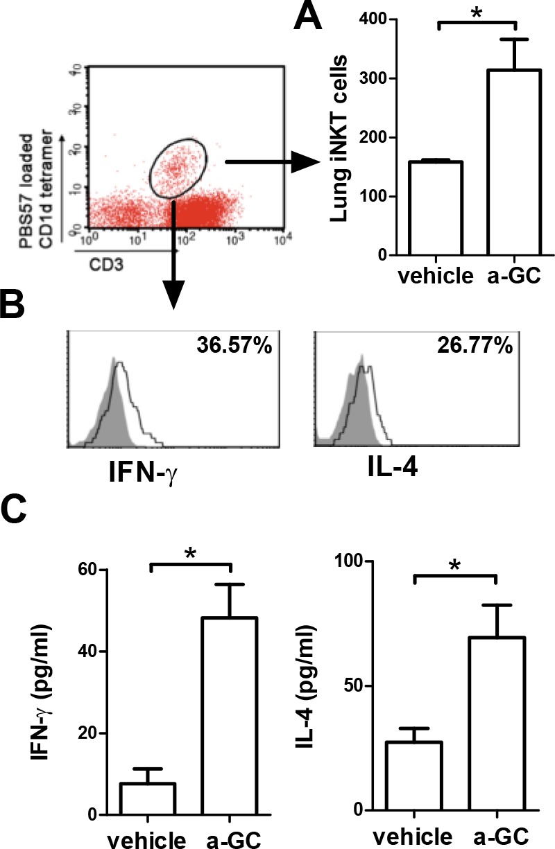
Mice were intranasally administered with vehicle or α-GalCer. Two hours later, BALF was collected. (A) The absolute numbers of iNKT (CD3+PBS57 loaded CD1d tetramer+) cells were measured. (B) IFN-γ and IL-4 expression from iNKT cells was assayed by intracellular staining with anti-mouse IFN-γ and IL-4 Abs. The shaded area is an isotype control; the open area reflects cytokine expression. The percentage of IFN-γ and IL-4-producing iNKT cells were shown. (C) IL-4 and IFN-γ levels in the BALF were measured by ELISA. a-GC, α-GalCer. n = 5 mice for each group. *, p<0.05 using Mann-Whitney U test.