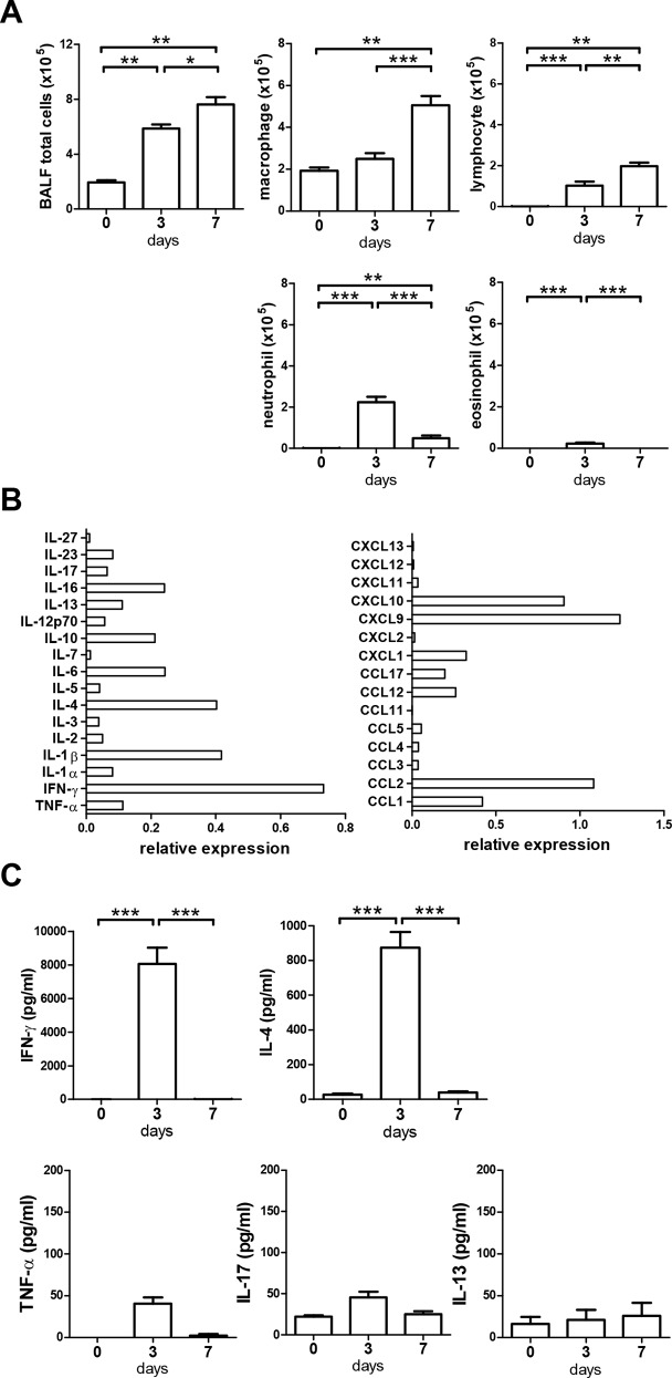
Fig 2. Intranasal α-GalCer administration induced acute inflammation in the lung.
Mice were intranasally administered with vehicle or α-GalCer. BALF was collected at days 0, 3 and 7. (A) BALF total cells and the absolute numbers of macrophages, lymphocytes, neutrophils, and eosinophils were measured at the indicated time points. (B) The relative expressions of cytokines and chemokines compared to positive control (set to a value of 1) of the assay kit in BALF at 3 days after α-GalCer administration of mice were determined by a dot blot immunoassay. Samples were pooled from 5–8 mice in two independent experiments. (C) Cytokines IFN-γ, IL-4, TNF-α, IL-17, and IL-13 in the BALF were assayed by ELISA. n = 5–8 mice for each group. *, p<0.05; **, p<0.01; ***, p<0.001 using Kruskal-Wallis one-way ANOVA followed Dunn's post test.

