
Fig 4. The presence of emphysema, mucus production and fibrosis in mice with repeated intranasal administration of α-GalCer.
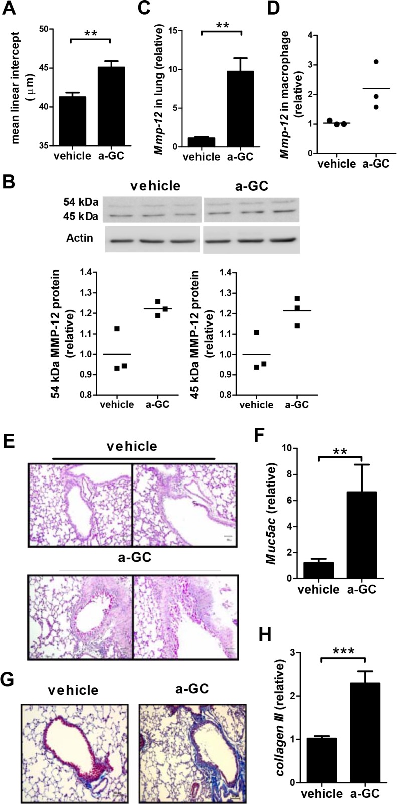
Mice were intranasally administered with α-GalCer once a week for 6 weeks and histopathological changes were examined 2 weeks after the last administration. (A) The mean linear intercept of the alveolar septa was measured. (B) The upper panels show the western blot results of 54 kDa and 45 kDa MMP12 protein expression in the lung and summarized in the lower panels. Actin was used as an internal control. Each symbol represents an individual mouse and the horizontal lines represent the means. (C) The expression of Mmp-12 in lung tissues was analyzed by quantitative RT-PCR. (D) The expression of Mmp-12 in lung macrophages was measured by quantitative RT-PCR. Each symbol represents cells pooled from 2–3 mice and the horizontal lines represent the means of three symbols. (E) Representative sections of the lung stained with PAS for analysis of mucus-containing cells. (200x magnification). (F) The expression of Muc5ac in lung tissues was analyzed by quantitative RT-PCR. (G) Representative results of Massion’s trichrome staining of lung sections (200x magnification, blue color). (H) The expression of collagen III in lung tissues was evaluated by quantitative RT-PCR. n = 9–14 mice for each group. a-GC, α-GalCer. **, p<0.01; ***, p<0.001 using Mann-Whitney U test.