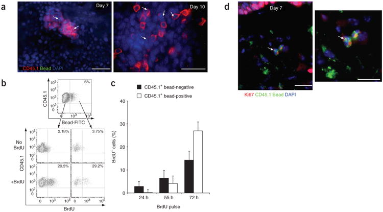
Figure 4.

Bead-positive Gr-1hi monocytes differentiate into dermal macrophages and epidermal LCs in inflamed skin. (a) Epidermal sheets stained for CD45.1 (red) and DAPI (blue) at 7 d and 10 d after ultraviolet irradiation. Fluorescein isothiocyanate–positive beads are bright green dots (arrows). Scale bars, 10 μm. Images are representative of at least four separate experiments. (b) Percent BrdU+ cells in recruited CD45.1+ bead-negative or CD45.1+ bead-positive populations in the epidermis after a 72-hour pulse of BrdU. Data are representative of three separate experiments. (c) Percent CD45.1+ bead-negative BrdU+ cells and CD45.1+ bead-positive BrdU+ cells after BrdU pulses of varying times. Data are one representative experiment (n = 3). (d) Frozen sections of skin stained for Ki67 (red), CD45.1 (green) and DAPI (blue), 7 d after ultraviolet treatment. Right, higher magnification. Fluorescein isothiocyanate–positive beads are bright green dots (arrows). Scale bars, 10 μm. Images are representative of two separate experiments.
